Table of Contents
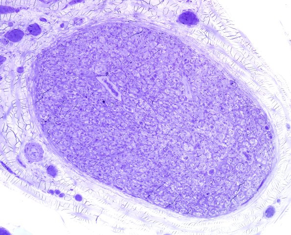
Washington University Experience | NEOPLASMS - CRANIAL AND PARASPINAL NERVEs | Perineurioma, Intraneural | 2E1 Perineurioma (Case 2) Plastic 5.jpg
2E1-3 One micron-thick, toluidine blue stained sections of the plastic embedded right sural nerve biopsy show marked loss of normally myelinated large and small axons, an increase in cellular processes, and highly abnormal intrafascicular architecture. Only scattered, thinly myelinated axons of small caliber remain. Numerous thick walled concentric whorls mimic onion bulbs but have thicker processes.

