Table of Contents
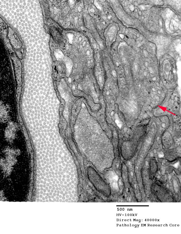
Washington University Experience | NEOPLASMS - CRANIAL AND PARASPINAL NERVEs | Perineurioma, Intraneural | 2F3 Perineurioma (Case 2) EM 057 copy - Copy
Higher magnification of the area marked by an arrow in image #2F2 (electron micrograph). The perineurial processes in this case contain numerous micropinocytotic vesicles (arrow) (electron micrograph)

