Table of Contents
Washington University Experience | NEOPLASMS - CRANIAL AND PARASPINAL NERVEs | Perineurioma, Intraneural | 3H5 Perineurioma (Case 3) Tumor_039 - Copy
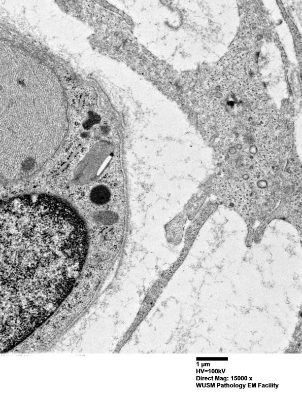
A portion of the image #3H4 at higher magnification shows a Schwann cell on the left with an intact continuous basement membrane versus the patchy and coarse perineurial processes on the right side of the image. (electron micrograph)

