Table of Contents
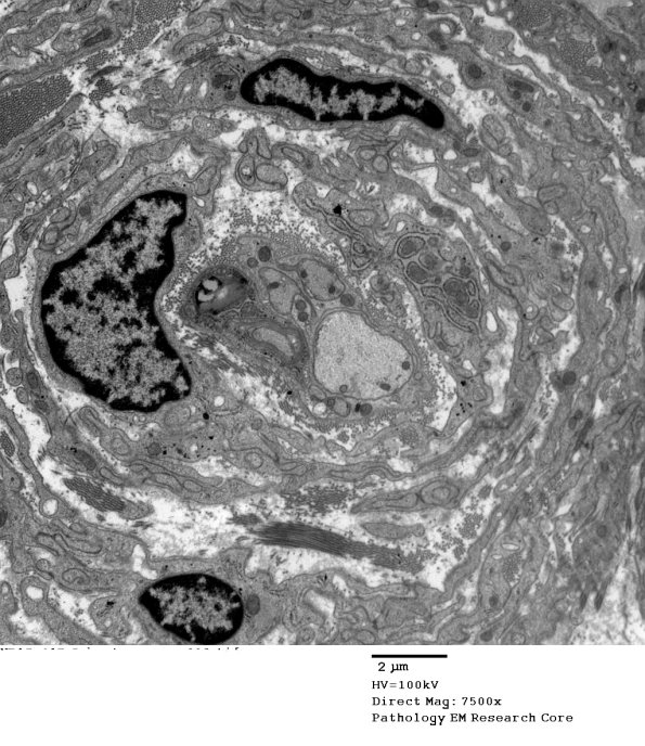
Washington University Experience | NEOPLASMS - CRANIAL AND PARASPINAL NERVEs | Perineurioma, Intraneural | 4F1 Perineurioma (Case 4) EM 003 - Copy
Electron microscopic evaluation of this nerve biopsy demonstrates decreased axonal density, likely due to expansion and infiltration by a cellular process that forms frequent whorls. Some whorls contain central axons often with thinned myelin as in this case. The cell types that form whorls demonstrate elongated tapered nuclei and long thin cytoplasmic processes with pinocytotic vesicles. (electron micrograph)

