Table of Contents
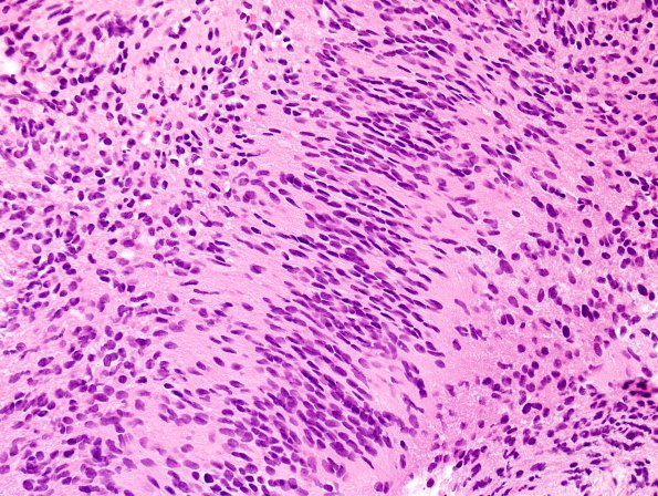
Washington University Experience | NEOPLASMS - CRANIAL AND PARASPINAL NERVEs | Schwannoma | 21B1 Schwannoma, 12yo girl (Case 21) H&E 3.jpg
21B1,2 Sections of the resected intradural extramedullary lesion show a schwannoma with classic as well as somewhat unusual histologic features. The majority is made up of solid Antoni A areas, but loosely textured Antoni B areas are present and there are well-formed Verocay bodies. Hyalinized vessels are present and there is an area suggestive of tumor capsule. A nerve root lies adjacent to the neoplastic outgrowth, which protrudes rather than growing along nerve fascicles as seen in neurofibroma. A few foci show hypercellularity, but these are not widespread.

