Table of Contents
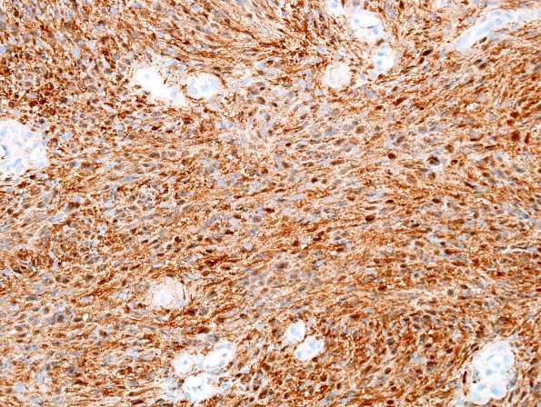
Washington University Experience | NEOPLASMS - CRANIAL AND PARASPINAL NERVEs | Schwannoma | 21C Schwannoma, 12yo girl (Case 21) S100 1.jpg
There is strong and diffuse S-100 immunoreactivity which stains the tumor cell cytoplasm as well as some nuclei. ---- Not shown: Neurofilament shows no entrapped axon fragments within the tumor mass. Glial fibrillary acidic protein is negative. Progesterone receptor and epithelial membrane antigen, markers of meningioma, are negative. The proliferation marker Ki-67 shows an overall low rate of proliferation. ---- Comment: This neoplasm predominantly has classic architectural, cytologic and immunohistochemical features of a well differentiated schwannoma. An unusual finding is the presence of necrosis, but in a neoplasm without atypia or mitotic activity, the presence of necrosis does not per se indicate aggressive behavior. Abnormal, fibrotic tumor vasculature may have been compromised by compression during growth.

