Table of Contents
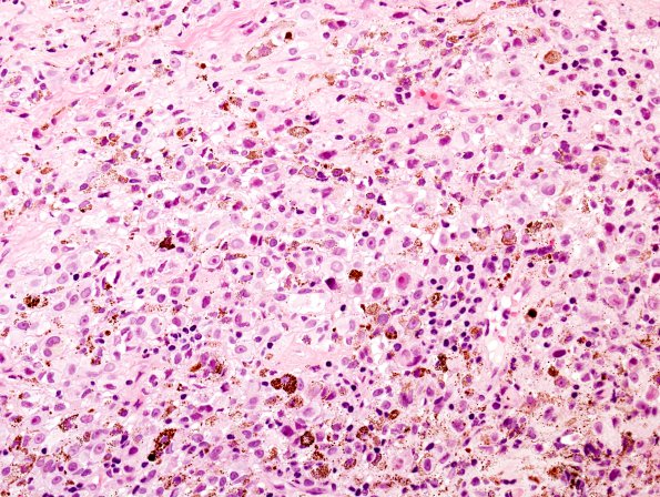
Washington University Experience | NEOPLASMS - CRANIAL AND PARASPINAL NERVEs | Schwannoma | 23A1 Schwannoma, melanotic (Case 23) H&E 1
Case 23 History ---- The patient is a 62-year-old man with a history of congestive heart failure, requiring pacemaker placement, chronic renal failure, and restless leg syndrome diagnosed 2 years prior, who has experienced worsening weakness and spasticity in his right leg for ten weeks, and has recently developed frank urinary incontinence. Non-contrast computed tomography reveals a small, apparently exophytic lesion of the spinal cord at level T10, and no evidence of other tumors. Intraoperatively, the lesion appears pigmented (black) and had a somewhat diffuse distribution. The patient has no history of melanoma; a keratoacanthoma was removed from his face and reviewed. Operative procedure: T10-11 laminectomy.
23A1,2 This is a pigmented neoplasm composed of tumor cells with variably spindled and epithelioid morphology, arranged in fascicles and sheets. A large subset of tumor cells is pigmented.

