Table of Contents
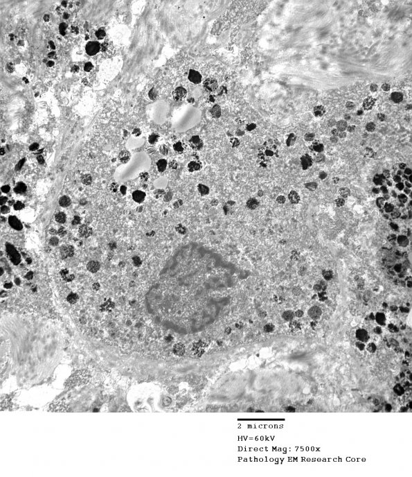
Washington University Experience | NEOPLASMS - CRANIAL AND PARASPINAL NERVEs | Schwannoma | 23F2 Schwannoma, melanotic (Case 23) EM 6 - Copy
Electron microscopic examination shows intracellular cytoplasmic melanosomes of various stages of maturation within the tumor cells. The tumor cell borders show a pattern of lamellar electron density and lucency consistent with basal lamina.

