Table of Contents
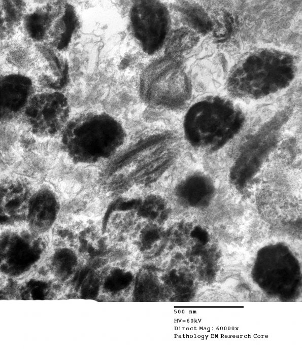
Washington University Experience | NEOPLASMS - CRANIAL AND PARASPINAL NERVEs | Schwannoma | 23F4 Schwannoma, melanotic (Case 23) EM 1 - Copy
Electron microscopic examination shows intracellular cytoplasmic melanosomes of various stages of maturation within the tumor cells. The tumor cell borders show a pattern of lamellar electron density and lucency consistent with basal lamina. ---- Not shown: There is strong cytoplasmic reactivity for intermediate filament vimentin in all tumor cells. Reactivity for S-100 protein, and for more specific melanotic markers, e.g. HMB-45 is present in the majority of tumor cells. No reactivity for epithelial membrane antigen (EMA) is observed. These features are consistent with melanotic schwannoma. ---- Comment: Given the anatomic location of this lesion, the histological pattern of variably epithelioid and spindled melanotic tumor cells suggests a differential diagnosis that includes melanoma, melanocytoma, and melanotic (pigmented) schwannoma. In this anatomic location, these three entities may be difficult to distinguish on the basis of routine histology. Although malignant melanoma usually demonstrates a high proliferative index and often atypical mitotic figures, exceptions exist. These three entities also share very similar immunoreactivity profiles, each at least potentially showing reactivity for classical 'melanoma markers' S-100, HMB-45 and Melan A. However, the presence of a basal lamina in the periphery of each cell, observable ultrastructurally, and by immunostaining with an antibody for collagen IV, has been reported to allow differentiation of melanotic schwannoma from the other two entities.

