Table of Contents
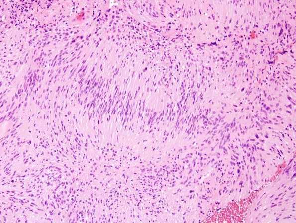
Washington University Experience | NEOPLASMS - CRANIAL AND PARASPINAL NERVEs | Schwannoma | 24A2 Schwannoma, myxoid (Case 24) H&E 5.jpg
Case 24 History ---- The patient is a 44 year old man who developed a limp while walking 4 years prior to admission. His symptoms progressed to urinary, bowel, and sexual dysfunctions. An MRI of his spine at an outside institution showed an intradural homogeneously enhancing mass at the level of the conus. Operative procedure: Thoracolumbar laminectomy for excisional biopsy of intradural lesion. ---- 24A1-4 Sections show an encapsulated mass consisting of neoplastic spindle-shaped cells forming short fascicles. The neoplasm contains areas of myxoid change. The neoplastic cells have predominantly elongated ovoid nuclei and a 'stretched' appearance. In addition cells with large plump nuclei with irregular nuclear contours are also present, consistent with so-called ancient (degenerative) change. Mitoses are rare (<1/10HPF). The neoplasm contains focal areas with well-defined Verocay body formation.

