Table of Contents
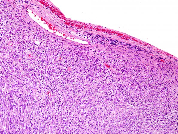
Washington University Experience | NEOPLASMS - CRANIAL AND PARASPINAL NERVEs | Schwannoma - Cellular | 2A1 Schwannoma, cellular (Case 2) H&E 2
Case 2 History ---- The patient is a 35-year-old woman with known neurofibromatosis type 2 (NF2) who on serial imaging was noted to have rapid progression of a large intradural extramedullary lesion at T12-L1 with significant cord compression. Operative procedure: T12-L1 laminectomy for excisional biopsy of intradural extramedullary lesion. ---- 2A1-3 Sections of the resected spinal mass show a relatively well-encapsulated spindle cell neoplasm arranged in variably oriented and sized fascicles. Verocay bodies are rare but present. Varying degrees of vascular hyalinization are seen. Regions that display significant crowding with nuclear overlap are accompanied by nuclear hyperchromasia and increased nuclear atypia with prominent nucleoli. The maximum mitotic rate is 11 mitoses/10HPF. Necrosis is absent. (H&E)

