Table of Contents
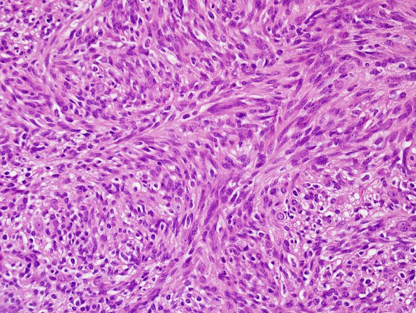
Washington University Experience | NEOPLASMS - CRANIAL AND PARASPINAL NERVEs | Schwannoma - Cellular | 2A3 Schwannoma, cellular (Case 2) H&E 5
Sections of the resected spinal mass show a relatively well-encapsulated spindle cell neoplasm arranged in variably oriented and sized fascicles. Verocay bodies are rare but present. Varying degrees of vascular hyalinization are seen. Regions that display significant crowding with nuclear overlap are accompanied by nuclear hyperchromasia and increased nuclear atypia with prominent nucleoli.

