Table of Contents
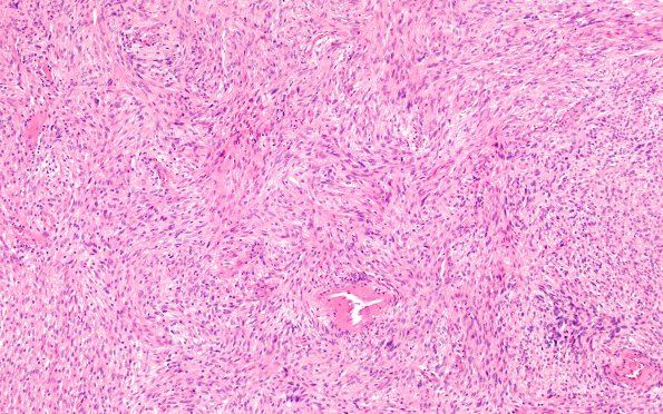
Washington University Experience | NEOPLASMS - CRANIAL AND PARASPINAL NERVEs | Schwannoma - Cellular | 3A1 Schwannoma, cellular (Case 3) H&E 10X 2
Case 3 History ---- The patient is a 33 year old female. Clinical history and diagnosis: Intradural/paraspinal mass status post posterolateral spine fusion. Operative procedure: Resection of retroperitoneal mass (right) and spinal fusion at L1 and L2 with bone graft. ---- 3A1-3 Sections of the specimens labeled retroperitoneal mass, paracaval tumor mass, and tumor of the left foramen show a cellular schwannoma. The tumor is composed predominantly of hypercellular (Antony A) areas populated by bland spindled cells with definitive nuclear palisading and occasional Verocay bodies. Rare large, hyperchromatic, irregular nuclei are noted, representing degeneration. Only rare mitotic figures are identified.

