Table of Contents
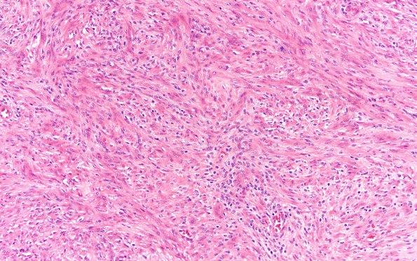
Washington University Experience | NEOPLASMS - CRANIAL AND PARASPINAL NERVEs | Schwannoma - Cellular | 5A2 Schwannoma, cellular (Case 5) H&E 20X 2
5A2,3 Microscopic examination of the "tumor" biopsy material shows a cellular schwannoma. The tumor consists largely of densely cellular tissue with a few small foci of loosely distributed tumor cells. The tumor cells have elongated mildly hyperchromatic and slightly pleomorphic nuclei with nucleoli of varying size. These are associated with elongated eosinophilic cytoplasm. Rare mitoses are identified. No necrosis is seen. In some areas tumor cells form fascicles and herringbone growth patterns. At the periphery, the tumor is encapsulated. Many lymphocytes are present in and surrounding the capsule. Fragments of peripheral nerve are also identified adjacent to the capsule.

