Table of Contents
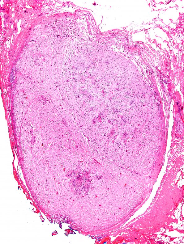
Washington University Experience | NEOPLASMS - CRANIAL AND PARASPINAL NERVEs | Schwannoma - Plexiform | 1B2 Schwannoma, plexiform, NF2 patient (Case 1) H&E 11
1B2,3 Individual neoplastic fascicles are expanded by an infiltrate of diffuse spindled cells with alternating areas of hypercellularity (including Verocay bodies) and hypocellularity (Antoni A and Antoni B areas). The nodular appearance is consistent with multiple individual fascicles resulting in a plexiform Schwannoma. Small tumorlets are seen within the endoneurium of multiple fascicles, with or without endoneurial expansion. The tumor cell nuclei in the main tumor mass are spindled and have only occasional pleomorphic forms. Mitotic rate is variable but overall is low with 2 mitoses/10HPF found focally.

