Table of Contents
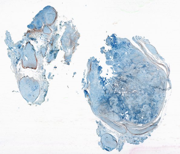
Washington University Experience | NEOPLASMS - CRANIAL AND PARASPINAL NERVEs | Schwannoma - Plexiform | 1G Schwannoma, plexiform, NF2 patient (Case 1) 1 EMA WM
Epithelial membrane antigen (EMA) is negative (evidence against a neoplastic perineurial component as might be seen in perineurioma. In addition, schwannoma expanded fascicles are easily seen outlined by EMA staining of the perineurium. The largest nodule has a few preserved residual elements. ---- Not shown: Ki-67 staining in the tumor is highly variable but elevated focally to 12.1%. The tumorlets have much fewer Ki-67 positive cells. ---- Comment #1: This patient had several paraspinal schwannomas resected over the years: 1) A routine schwannoma at L4 in 2012 with low proliferative activity (2 mitoses/10HPF) and represents the plexiform pattern shown here. ---- 2) Three years later she had a T12 cellular schwannoma with increased mitotic rate (11 mitoses/10 HPF) (Cellular Schwannoma case 2 in this atlas). Support for a schwannian neoplasm was provided by strong, diffuse immunoreactivity for the transcription factor SOX10. Diffuse SOX 10 positivity of tumor cells is support for the diagnosis of cellular schwannoma when compared to malignant peripheral nerve sheath tumors (PMID: 25189642). (SOX10 IHC). ---- 3&4) In 2020 surgeries for T4 and T10-11 showed routine schwannomas. ---- Comment Presence of high cellularity, increased mitotic activity, patchy p53 immunoreactivity, and partial loss of S100 protein expression are somewhat worrisome findings in her second neoplasm but given its overall small size and a 'schwannoma' phenotype, we and colleagues favored all these changes to be within the spectrum of cellular schwannoma albeit with slightly elevated mitotic activity. The morphological and immunohistochemical features are consistent with a schwannoma (WHO Grade I) in a patient with NF2.

