Table of Contents
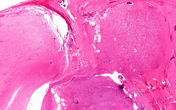
Washington University Experience | NEOPLASMS - CRANIAL AND PARASPINAL NERVEs | Schwannoma - Plexiform | 3A3 Schwannoma, plexiform (Case 3) B2 H&E 2X
The tumor mass show expanded fascicles. Sections of the specimens show a spindle cell neoplasm with associated non-neoplastic nerve fascicles. This tumor is composed of one main large tumor nodule with bimorphic growth patterns (cellular or Antoni A and loose or Antoni B regions), and several smaller nodules composed of expanded nerve fascicles with intrafascicular growth of neoplastic spindle cells with features similar to those in the main nodule. These structures represent expansion/presence of tumor into contiguous nerves and impart a plexiform appearance to the tumor. Mitoses are difficult to find.

