Table of Contents
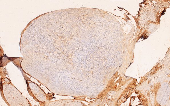
Washington University Experience | NEOPLASMS - CRANIAL AND PARASPINAL NERVEs | Schwannoma - Plexiform | 3D2 Schwannoma, plexiform (Case 3) B2 EMA 4X 3
EMA immunohistochemistry may highlight the plexiform nature of the neoplasm by labeling the perineurium of individual fascicles and demonstrating their expanded size and contents. If fascicular nodules are too large the small amount of residual staining in the walls may be limited. ---- Not shown: Tumor cells produce basal lamina highlighted by collagen IV immunoreactivity, but stain negative for EMA. Taken together, both morphologic features and immunoprofiles of this tumor support the diagnosis of plexiform schwannoma which has not been shown convincingly in most cases to be associated with neurofibromatosis type I or II.

