Table of Contents
Washington University Experience | NEOPLASMS (HEMATOLYMPHOID) | Langerhans Cell Histiocytosis (LCH) | 11A LCH (Case 11 CT skull series 1 - Copy
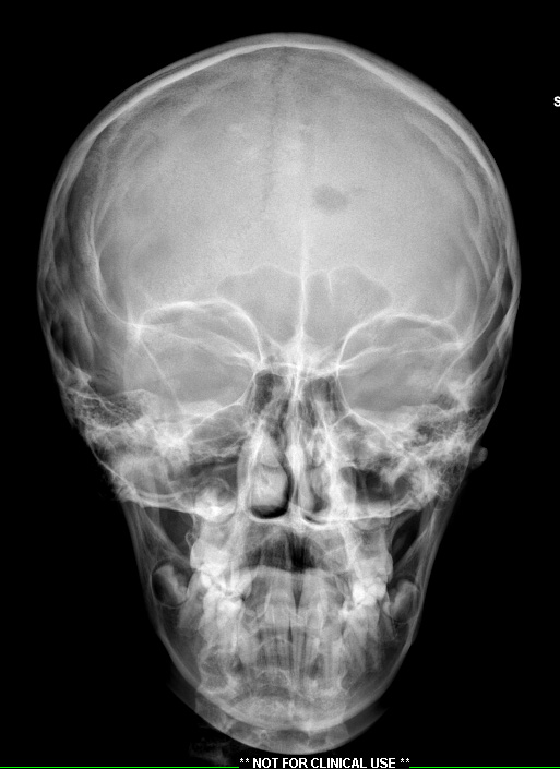
Case 11 History ---- The patient was a 10-year-old boy with a skull lesion. ----
11A CT scan showed this discrete lesion in the skull. There is enhancement of the subcutaneous/periosteal component over the left frontal bone. Additional MRI examination showed an 8mm T1 isointense and T2 hyperintense enhancing lesion in the anterior left frontal bone near the midline. Operative procedure: Left frontal skull lesion biopsy.

