Table of Contents
Washington University Experience | NEOPLASMS (HEMATOLYMPHOID) | Langerhans Cell Histiocytosis (LCH) | 11B LCH (Case 11) H&E 1
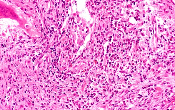
H&E-stained permanent sections of the biopsied left frontal skull lesion showed fragments of soft tissue and bony trabeculae involved by patches of markedly inflamed tissue and clusters of atypical histocytes. These nodular and diffuse foci of chronic inflammation are composed of predominantly mature looking lymphocytes as well as scattered eosinophils. The admixed histiocytes in these clusters have moderate nuclear pleomorphism, reniform nuclei, and abundant eosinophilic cytoplasm. Occasional multinucleated reactive histiocytes are also seen. The soft tissue consists mostly of fibrovascular tissue and myofibroblastic proliferation likely due to intense inflammation.

