Table of Contents
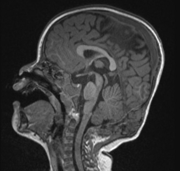
Washington University Experience | NEOPLASMS (HEMATOLYMPHOID) | Langerhans Cell Histiocytosis (LCH) | 12A1 LCH (Case 12) T1 MPRAGE no C - Copy
Case 12 History ---- The patient was a 21-month-old boy with no significant past medical history who was noted to have a growing occipital skull lesion present for 2 months prior to workup. ---- 12A1,2 MRI demonstrated a 5.0 x 4.6 x 2.0 cm soft tissue mass arising from the midline occiput that abuts the dura overlying the posterior cerebellum, inferior posterior occipital cortex and confluence of the venous sinuses. Operative procedure: Biopsy and curettage of occipital lesion.

