Table of Contents
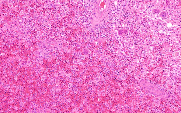
Washington University Experience | NEOPLASMS (HEMATOLYMPHOID) | Langerhans Cell Histiocytosis (LCH) | 12B1 LCH (Case 12) H&E 4
12B1,2 H&E stained sections show a lesion composed of cells with irregular to folded nuclei with significant nuclear grooves, fine granular or vesicular chromatin, distinct nucleoli and moderate to abundant pale eosinophilic cytoplasm. Frequent admixed eosinophils are seen. There are also multiple giant cells that resembling osteoclasts in appearance, hemosiderin-laden macrophages that are positive for iron stain, and hyalinized collagen fibers are focally numerous in the background. The lesion appears to be infiltrating skeletal muscle.

