Table of Contents
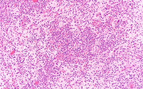
Washington University Experience | NEOPLASMS (HEMATOLYMPHOID) | Langerhans Cell Histiocytosis (LCH) | 13A1 LCH (Case 13) H&E 20X
Case 13 History (Same patient as Case 12) ---- The patient was a 2-year-old boy with history of Langerhans cell histiocytosis (same cases as Case 12 previous) that presented as an occipital mass, s/p resection in 8/2020. Due to the occipital wound drainage, an occipital/suboccipital exploration was performed with intraoperative findings concerning for recurrence. Culture showed growth of MRSA. Operative procedure: Wound exploration and resection of occipital/suboccipital epidural lesion. ---- 13A1,2 H&E stained sections show the neoplasm resected 4 months after Case 12 composed of loosely cohesive sheets of histiocytic cells demonstrating round to oval to crescent nuclei, conspicuous nuclear grooves, fine granular chromatin, scattered nucleoli and scant to moderate amount of eosinophilic cytoplasm. Numerous eosinophils, neutrophils, lymphocytes, plasma cells as well as a few multinucleate giant cells are variably admixed. Mitoses are scattered. Surface ulceration with areas of necrosis, including infarcted/necrotic fragments of epidermis, granulation tissue, and scattered hemosiderin were also identified.

