Table of Contents
Washington University Experience | NEOPLASMS (HEMATOLYMPHOID) | Langerhans Cell Histiocytosis (LCH) | 14B1 LCH (Case 14) 2.jpg
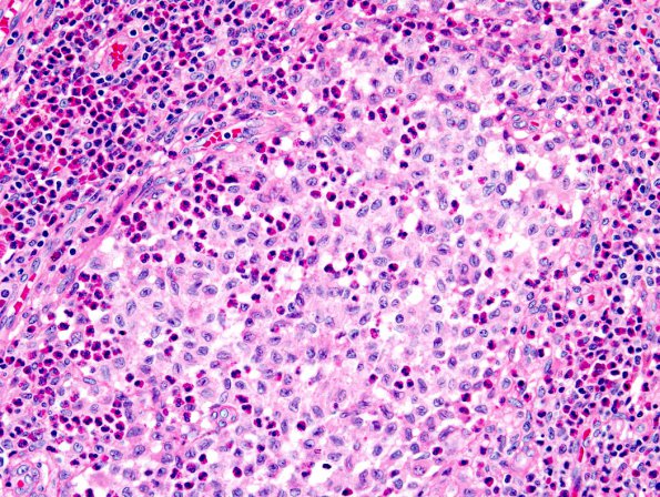
14B1-3 Sections of the left frontal mass and bone show sheets of loosely cohesive epithelioid cells with irregular, grooved vesicular nuclei and moderate amounts of amphophilic to eosinophilic cytoplasm. There are foci of abundant eosinophils and scattered reactive multinucleated giant cells. The lesion is surrounded by a thick, fibrous capsule consistent with dura or periosteum.

