Table of Contents
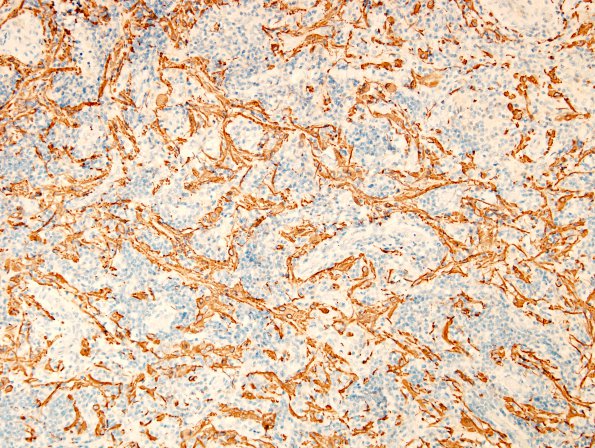
Washington University Experience | NEOPLASMS (HEMATOLYMPHOID) | Langerhans Cell Histiocytosis (LCH) | 16E LCH (Case 16) GFAP
Brain invasion of the process is demonstrated by GFAP immunohistochemistry (GFAP IHC). ---- Ancillary findings (not shown): Synaptophysin stains highlight background glial processes and neuropil, respectively. A NeuN stain highlights the absence of significant numbers of neurons. These features, together with the radiographic features of an isolated, expansile, lytic lesion of the vertebral body is consistent with Langerhans cell histiocytosis (LCH).

