Table of Contents
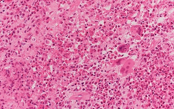
Washington University Experience | NEOPLASMS (HEMATOLYMPHOID) | Langerhans Cell Histiocytosis (LCH) | 19B1 LLCH (Case 19) H&E 40X 2
19B1,2 Microscopic sections of the skull lesion show a cellular histiocytic neoplasm with numerous associated eosinophils. Neoplastic cells have abundant eosinophilic cytoplasm with ovoid to reniform nuclei with frequent longitudinal nuclear grooves and intranuclear inclusions. Numerous osteoclast-like giant cells are also present. In addition to pronounced eosinophilia, there is a scattered mixed inflammatory background that includes neutrophils and lymphocytes.

