Table of Contents
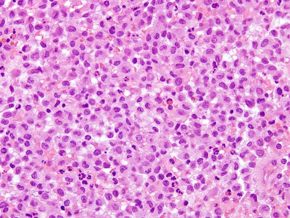
Washington University Experience | NEOPLASMS (HEMATOLYMPHOID) | Langerhans Cell Histiocytosis (LCH) | 1A1 LCH (Case 1) H&E 5
Case 1 History The patient was a one year old boy with a right temporal skull lesion. Operative procedure: Excision of right temporal skull lesion. ---- 1A1,2 The neurosurgical specimen consists of an infiltrate composed by cells with eccentric, ovoid, reniform nuclei with linear grooves and inconspicuous nucleoli, consistent with Langerhans cells, as well as macrophages, lymphocytes, plasma cells and eosinophils. There are scattered multinucleated giant cells.

