Table of Contents
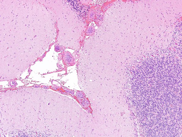
Washington University Experience | NEOPLASMS (HEMATOLYMPHOID) | Lymphoma, Intravascular Transforms to Primary | 1B1 Lymphoma, intravascular (Case 1) H&E 10.jpg
1B1-3 Sections from the left cerebellar cortex show superficial cerebellar cortex and leptomeninges involved by a high grade intravascular malignancy. Most of the small to intermediate sized leptomeningeal and superficial cortical blood vessels are filled with discohesive malignant cells with scant cytoplasm, oval nuclei with irregular nuclear contours, vesicular chromatin and prominent nucleoli. Mitoses are numerous. The malignant cells occlude most of the involved blood vessels, but appear to be largely confined to the intravascular spaces and do not infiltrate brain parenchyma. The cerebellar cortex otherwise shows several microinfarcts at different stages of resolution. There is eosinophilic neuronal necrosis of the Purkinje cells and patchy Bergmann gliosis, suggesting recent and prior ischemic damages.

