Table of Contents
Washington University Experience | NEOPLASMS (HEMATOLYMPHOID) | Lymphoma, PTLD | 1B1 Lymphoma, B-cell (PTLD, Case 1) _1 copy
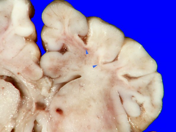
1B1-3 The brain was overall quite soft in consistency. Coronal sections of the cerebral hemispheres revealed indistinct demarcation between the gray and white matter, consistent with the age of the patient. The convolutional and central white matter are soft with focal discoloration. The cortex shows a few foci of ulegyria (arrowheads, 1B1) in the frontal lobes. The various nuclear components of the basal ganglia and thalami appear discolored (tan-brown). Sections of the cerebellum show atrophy and multiple foci of softening including the cortex.

