Table of Contents
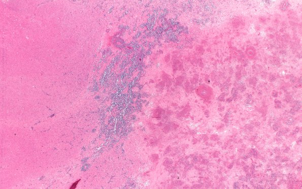
Washington University Experience | NEOPLASMS (HEMATOLYMPHOID) | Lymphoma, immune-compromised | 10B1 Lymphoma, (Case 10) H&E 2X
10B1,2 Microscopic examination of the right basal ganglia mass and the left caudate are similar in appearance with a large number of lymphoid cells arranged perivascularly and throughout the parenchyma. There is extensive necrosis with admixed reactive lymphocytes and astrocytes. The right frontal lesion is hemorrhagic secondary to the biopsy of the deeper lesion. Numerous multinucleated cells, indicative of HIV infection, are associated with the areas of lymphoma. Sections of the right frontal lobe lesion show some acute ischemic changes with eosinophilic neurons as well as hemorrhage, but no obvious lymphoid cells. Other areas of the cortex and brainstem appear normal without multinucleated HIV infected cells. Stains for fungus, a CMV immunostain, and a herpes immunostain are all negative.

