Table of Contents
Washington University Experience | NEOPLASMS (HEMATOLYMPHOID) | Lymphoma, immune-compromised | 7A1 Lymphoma, HIV, CMV (Case 7)
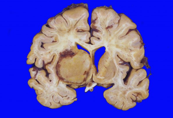
7A1-3 Coronal sections through the cerebral hemispheres demonstrate widening of the sulci and some enlargement of the left and right lateral ventricles consistent with cerebral atrophy. The left basal ganglia is effaced by a 5 cm mass of tan, brown necrotic tissue that extends from the anterior aspect of the left lateral ventricle caudally to the thalamus at the level of the substantia nigra. The mass focally extends laterally to the external capsule demonstrates a hyperemic lateral and inferior rim and areas of hemorrhage are also present laterally. A 1cm greatest dimension nodule of tissue that is identical in color and consistency is present in the right caudate nucleus head. There is evidence of CMV ependymitis/encephalitis.

