Table of Contents
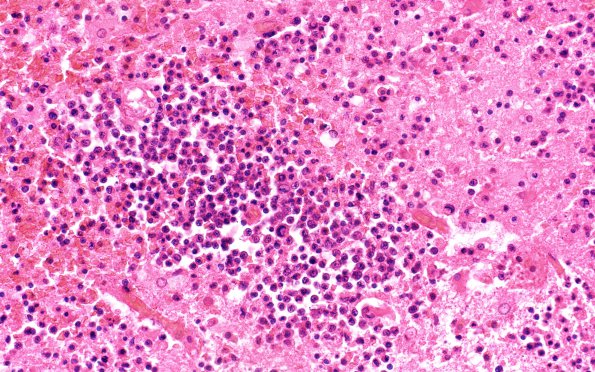
Washington University Experience | NEOPLASMS (HEMATOLYMPHOID) | Lymphoma, immune-compromised | 7B1 Lymphoma, HIV, CMV (Case 7) 40X
7B1,2 Sections of the left and right basal ganglia and left frontal-parietal lobe show malignant lymphoma. The neoplasm in all locations is largely necrotic and most of the viable neoplastic cells are present in the Virchow-Robin space of large and small blood vessels that surround the mass lesions. The neoplastic cells demonstrate slightly enlarged, hyperchromatic nuclei and scant cytoplasm. Mitotic figures are common.

