Table of Contents
Washington University Experience | NEOPLASMS (HEMATOLYMPHOID) | Lymphoma, immune-compromised | 8A1 Lymphoma, HIV patient (Case 8) 5
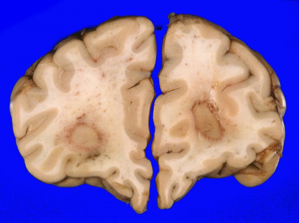
8A1-3 At autopsy serial coronal sections of the cerebral hemispheres at 1 cm intervals reveal numerous parenchymal lesions. Bilaterally in the frontal white matter there are 2.0 cm lesions with whitish-grey centers with a thin rim of surrounding hemorrhage. Similar appearing lesions are present in the left parietal cortex (1.0 cm), left temporal cortex (2.0 cm), and right parietal-temporal cortex (1.0 cm). In the right centrum semiovale there is a 3 x 3 x 2 cm firm, white lesion with surrounding hemorrhage. In addition, there is diffuse swelling in the left temporal lobe.

