Table of Contents
Washington University Experience | NEOPLASMS (HEMATOLYMPHOID) | Lymphoma, immune-compromised | 9A Lymphoma (Case 9) 2.jpg
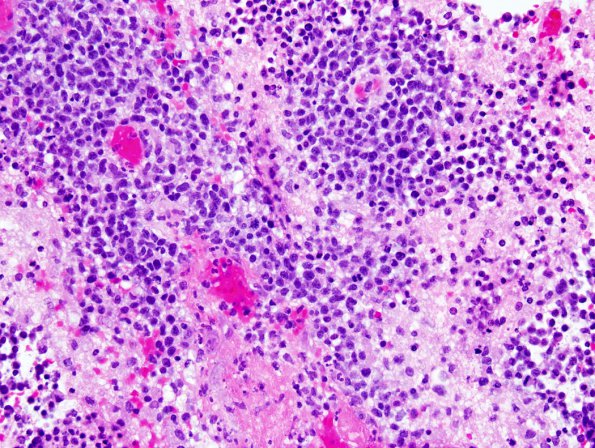
Case 9 History ---- The patient is a 54-year-old man who was HIV positive, and had a 6 x 6.7 x 6 cm left frontal lobe mass which is heterogeneous on T1 and T2 scans, shows irregular peripheral enhancement, signal loss on T2 star sequence, and no diffusion enhancement. The lesion extends into the corpus callosum anteriorly. Clinical diagnosis: Lymphoma more likely than high-grade glioma. Operative procedure: Biopsy of left frontal brain mass. ---- 9A Histologic sections of the left frontal tumor show an extensively necrotic neoplastic lesion comprised by large, abnormal lymphocytes with amphophilic cytoplasm, a high nuclear to cytoplasmic ratio, irregular nuclear contours, open chromatin, and prominent nucleoli. These atypical cells show abundant mitotic figures, and are arranged angiocentrically around abnormal-appearing benign blood vessels that have a plump endothelial lining. Subtly intermixed within these cuffs of larger atypical lymphocytes are infrequent, benign-appearing lymphocytes.

