Table of Contents
Washington University Experience | NEOPLASMS (HEMATOLYMPHOID) | Lymphoma, primary | 10A1 Lymphoma (Case 10) 3
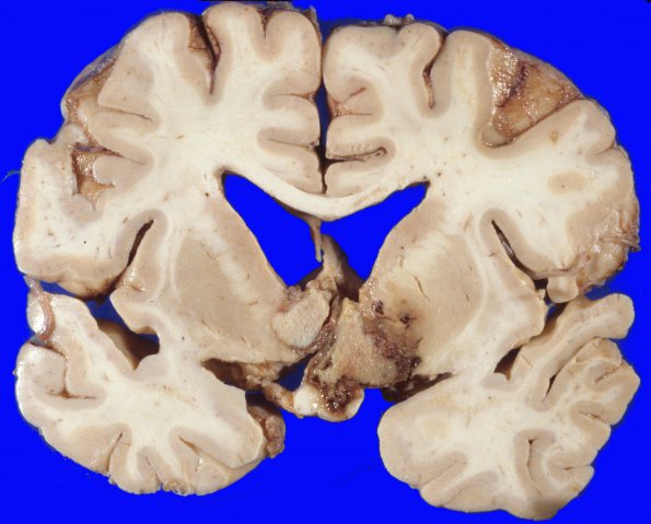
10A1-3 At autopsy the ventral surface of the brain is remarkable for elevation of the optic nerves and chiasm by an ill-defined greyish mass protruding from the region of the third ventricle. Coronal sections of the cerebral hemispheres show a large circumscribed granular partially necrotic mass occupying the right hypothalamus extending anteriorly to the level of the basal ganglia. There is also extension of the mass across the midline into the left caudate nucleus and also into the internal capsule of the chiasm. The mass involves the optic chiasm and the right optic tract. The caudal extension of the mass is to the mid-thalamus. The mass on the right extends into the globus pallidus and caudate nucleus. ---- 10A1-3 Coronal sections of the cerebrum and deep white matter shows prominent necrosis.

