Table of Contents
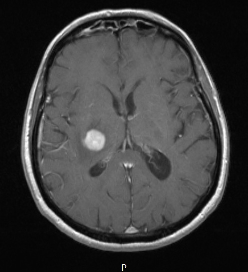
Washington University Experience | NEOPLASMS (HEMATOLYMPHOID) | Lymphoma, primary | 13A Lymphoma, primary CNS, B-cell (Case 13) T1W - Copy
Case 13 History ---- The patient is a 70 year old man who presented with progressive left-sided numbness in August of 2013. He initially presented in early 2012 with altered mental status and MRI workup from February through September 2012 showed multiple areas of T2/FLAIR hyperintensities with post-contrast enhancement throughout the white matter. A biopsy was attempted from one of the lesional areas in the left frontal lobe, but it was non-diagnostic. The lesions were responsive to steroid treatment in the past. Recent MRI in September 2013 showed interval enlargement of the right parietal and deep white/gray matter enhancing lesions with increasing vasogenic edema. Operative procedure: Stereotactic biopsy of right parietal lesion using Stealth navigation. ---- A mass lesion is well seen in this T-1 weighted contrast enhanced MRI scan.

