Table of Contents
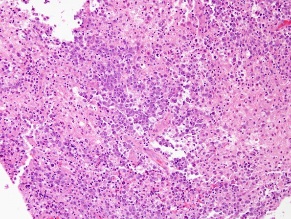
Washington University Experience | NEOPLASMS (HEMATOLYMPHOID) | Lymphoma, primary | 13B1 Lymphoma, primary CNS, B-cell Case 13) H&E 1.jpg
13B1,2 Hematoxylin and eosin stained sections show multiple fragments of hypercellular brain parenchyma involved variably by a malignant neoplasm arranged in solid sheet-like growth pattern along-with with some areas of diffuse infiltration. Some of the fragments have notable expansion of Virchow-Robin spaces by these malignant cells.

