Table of Contents
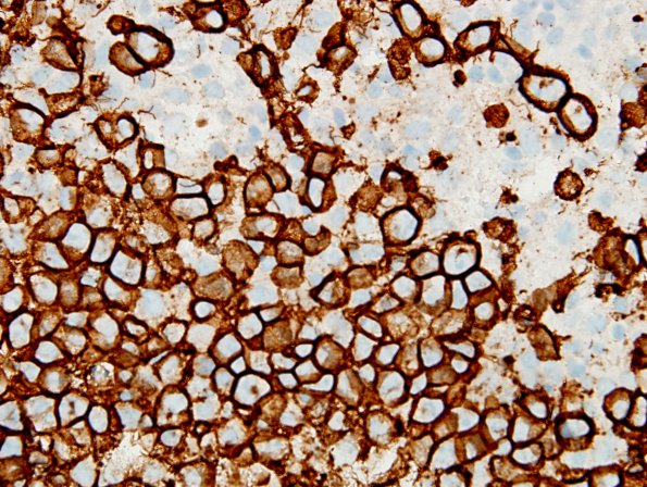
Washington University Experience | NEOPLASMS (HEMATOLYMPHOID) | Lymphoma, primary | 13C2 Lymphoma, primary CNS, B-cell (Case 13) CD20 60X 0 setting.jpg
Immunohistochemistry shows that the neoplastic cells are strongly and diffusely positive for the B-cell lymphocyte marker CD20. (CD20 IHC) ---- Other Immunohistochemistry (not shown): Tumor cells are negative for GFAP and synaptophysin although the former does highlight the presence of reactive astrocytes. Presence of scattered T-lymphocytes is supported by CD3 immunoreactivity. The proliferation marker Ki-67 stains a very large subset of tumor cell nuclei.

