Table of Contents
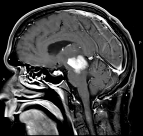
Washington University Experience | NEOPLASMS (HEMATOLYMPHOID) | Lymphoma, primary | 16A2 Lymphoma (Case 16) T1 W 2 - Copy
The second MRI shows the lesion in a sagittal cut. Other lesions are present in the right frontal lobe, left thalamus, left caudate and basal ganglia, left temporal lobe, and cerebellar hemisphere.

