Table of Contents
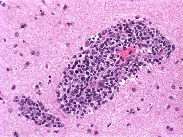
Washington University Experience | NEOPLASMS (HEMATOLYMPHOID) | Lymphoma, primary | 20A Lymphoma (Case 20) H&E 1
Case 20 History ---- The patient is a 53-year-old man with an enhancing lesion in the left frontoparietal area which shrank following steroid treatment. Clinical diagnosis: Glioblastoma or lymphoma. Operative procedure: Stereotactic biopsy. ---- 20A Sections of the specimen show fragments of brain parenchyma and densely-packed infiltrate of atypical lymphocytes with pleomorphic nuclei and scant cytoplasm. The cells range from intermediate to large size and have round to oval nuclei with smooth or irregular nuclear contours and one to multiple prominent nucleoli. Mitotic figures and apoptotic bodies are frequently identified.

