Table of Contents
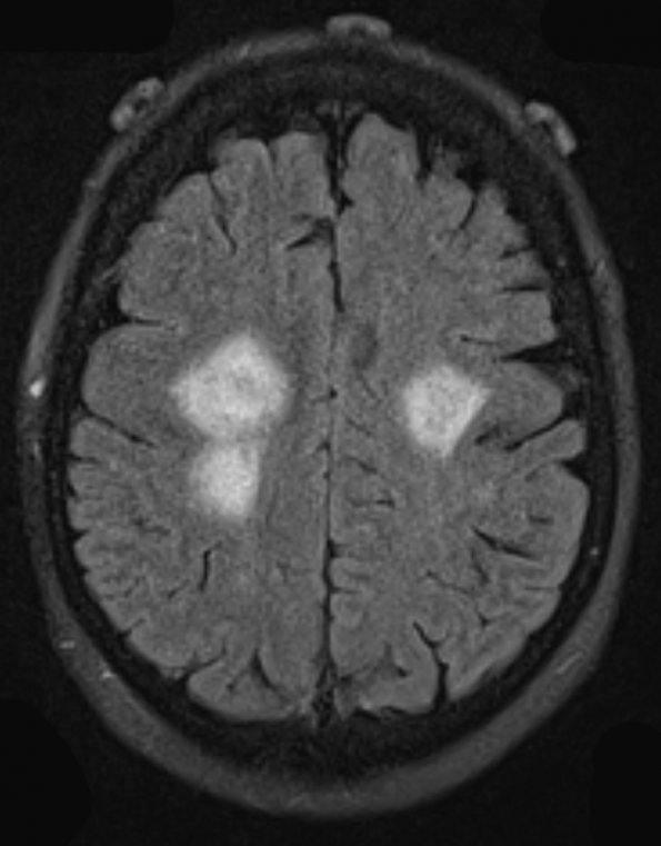
Washington University Experience | NEOPLASMS (HEMATOLYMPHOID) | Lymphoma, primary | 21A1 Lymphoma (Case 21) FLAIR 1 - Copy
Case 21 History ---- The patient is a 64 year old woman with recent facial, arm, and leg weakness. ---- 21A1,2 MRI shows multiple hyperintense lesions on FLAIR (21A1) and a T2-weighted scan with contrast (21A2) involving the right frontal lobe centrum semiovale and extending caudally through the posterior limb of the internal capsule and basal ganglia, into the right cerebral peduncle. Similar lesions are present in the subcortical left frontal lobe white matter just anterior to the central sulcus, and situated posteromedially along the frontal side of the central sulcus. Radiological differential diagnosis: Lymphoma, glioma, or demyelination. Operative procedure: Left frontal stereotactic brain biopsy.

