Table of Contents
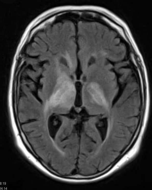
Washington University Experience | NEOPLASMS (HEMATOLYMPHOID) | Lymphoma, primary | 5A1 Lymphoma (Case 5) FLAIR 2 - Copy
Case 5 History ---- The patient is a 69 year old woman with several months of deteriorating neurological status, left-sided weakness, and left upper extremity hemiballismus. MRI of the brain shows large areas of signal abnormality within the white matter of both cerebral hemispheres. Lesions involve the periventricular white matter in the frontal lobes, body and splenium of the corpus callosum, and corticospinal tracts. There is also an enhancing nodule present within the right cerebral peduncle. Operative procedure: Right frontal burrhole and stereotactic needle biopsy. ---- 5A1-4 MRI studies: 5A1 Hyperintensity of T2 FLAIR elements, especially in the internal capsule.

