Table of Contents
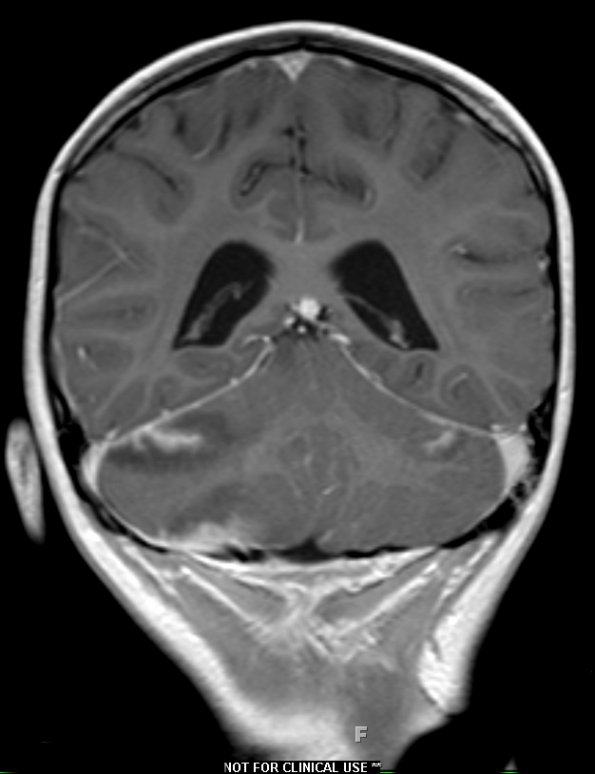
Washington University Experience | NEOPLASMS (HEMATOLYMPHOID) | Lymphoma, secondary | 1A1 Lymphoma, anaplastic large T cell, secondary (Case 1) T1 W - Copy - Copy
Case 1 History ---- The patient was a nine-year-old girl with history of anaplastic large cell lymphoma, status post chemotherapy, who recently presented with headache and neutropenic fever. CT and MRI show multiple areas of leptomeningeal enhancement and a posterior fossa space occupying mass lesion. Operative procedure: Craniotomy for evacuation of mass. ---- 1A1,2 Multiple nodular and linear branching enhancing lesions are seen throughout the cerebrum, involving the right frontal lobe, including the gyrus rectus, left temporal lobe, right greater than left cerebellar hemispheres, and vermis as seen on the T1-weighted with contrast scan (1A1) and high signal intensity on the T2-weighted with contrast scan (1A2). Edema is noted throughout the posterior two thirds of the right cerebellar hemisphere.

