Table of Contents
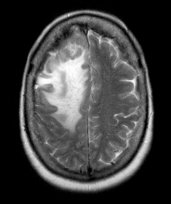
Washington University Experience | NEOPLASMS (HEMATOLYMPHOID) | Lymphoma, secondary | 3A1 Lymphoma, Secondary DLBCL to Dura (Case 3) MRI 4 - Copy - Copy
Case 3 History ---- The patient is a 67-year-old woman with a history of B-cell lymphoma, diagnosed from a neck biopsy as a diffuse large B-cell lymphoma, germinal center type (BCL6+, MUM1-). She presented within a year with a dural-based right frontal lesion. Operative procedure: Resection ---- 3A1,2 The dura based lesion is shown in FLAIR (with adjacent brain edema, 3A1) and in a T1-weighted contrast administered scan (3A2).

