Table of Contents
Washington University Experience | NEOPLASMS (HEMATOLYMPHOID) | Lymphoma, secondary | 6B1 Lymphoma, secondary (Case 6) H&E 3.jpg
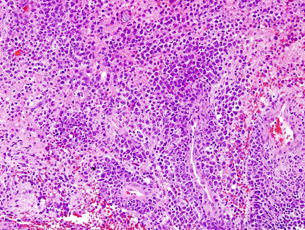
6B1,2 H&E stained biopsy material shows multiple small fragments of the neurosurgical biopsy extensively involved by a discohesive, infiltrative neoplasm distributed in angiocentric and sheeted patterns. Individual tumor cells have large, mildly irregular oval nuclei, coarsely granular chromatin, a few prominent nucleoli and minimal cytoplasm. Mitotic figures, apoptotic bodies and patches of necrosis or foamy macrophages are frequent. Adjacent brain parenchyma shows reactive astrocytosis.

