Table of Contents
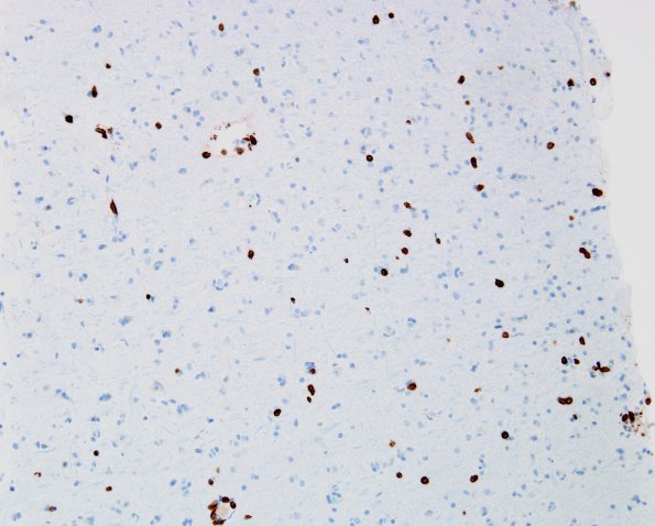
Washington University Experience | NEOPLASMS (HEMATOLYMPHOID) | Lymphomatosis cerebri | 1D2 Lymphomatosis cerebri (Case 1) CD3 20X
Immunohistochemical study of the same area of the biopsy (shown as 1D1-3) stained for CD3 (T cells, non-neoplastic) is shown at 20X magnification.

