Table of Contents
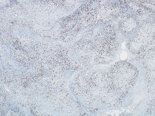
Washington University Experience | NEOPLASMS (MENINGIOMA) | Anaplastic | 11D Meningioma, anaplastic (Case 11) Ki67 2
11D A Ki-67 proliferation index is quite high throughout, reaching ~40.2% in more active areas. ---- Ancillary data (not shown): The abundance of chronic inflammatory cell infiltrate which is composed of lymphocytes and plasma cells is highlighted by CD45 and CD138 immunostains respectively with predominance of CD3 positive T lymphocyte population and a minor component of CD20 positive B-lymphocytes. Admixed histiocytic cells in the inflammatory infiltrate are reactive for CD68, and in-situ hybridization for kappa and lambda chains reveal a polyclonal plasma cell population. Presence of large areas of hyalinization is supported by a trichrome histochemical stain; these areas are negative for amyloid deposition by a thioflavin S stain. ---- FISH was performed utilizing probes against 1p32/14q32 and BCR/NF2. In this particular case, deletions of all markers were identified, consistent with losses involving 1p, 14q and 22q. Specifically, deletions of BCR and NF2 consistent with loss of 22q was noted in 90% of the 200 interphase nuclei examined and co-deletion of 1p32 and 14q32 was noted in 71% of the 200 interphase nuclei examined. These genetic alterations are often associated with higher grade meningiomas and aggressive behavior. ---- Comment: The overall findings are those of an anaplastic meningioma (WHO grade III) which is also brain invasive.

