Table of Contents
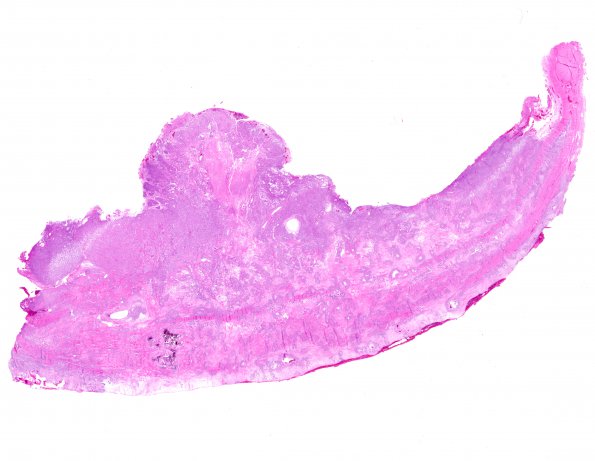
Washington University Experience | NEOPLASMS (MENINGIOMA) | Anaplastic | 12A1 Meningioma, Anaplastic (Case 12) H&E WM
Case 12 History ---- The patient was a 55 year-old man with recent seizure and an intracranial/extracranial frontal/parietal/temporal mass, dura based. Biopsy at outside hospital consistent with anaplastic meningioma. Mitotic figures were numerous with the count exceeding 20 mitoses/10 HPF focally. The workup of that consult case involved FISH that demonstrated deletions of 22q, 1p, and 14q. A diagnosis of anaplastic meningioma was made. ---- The next year additional surgery was performed. ---- 12A1-6 The tumor consisted of cords, sheets, and small nodules of atypical epithelioid cells with eosinophilic cytoplasm and prominent nucleoli. The tumor cells are surrounded and embedded within a highly collagenous matrix. Mitotic figures are easy to identify with up to 11 mitotic figures per 10 high-power fields focally. Brain invasion is also present.

