Table of Contents
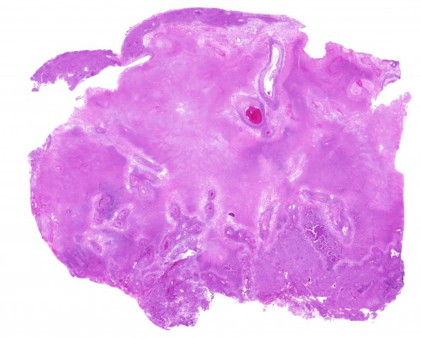
Washington University Experience | NEOPLASMS (MENINGIOMA) | Anaplastic | 13C2 Meningioma, anaplastic (Case 13) H&E whole mount 2
13C2-6 H&E stained sections of the resection material show a high grade meningioma with large, predominantly central, areas of geographic necrosis that comprise approximately 80% of the specimen. Areas of viable tissue with generally meningothelial features show marked hypercellularity, 'sheeted' architecture, foci of spontaneous necrosis, and focal 'small cell change.' Other areas that contribute less than 50% of the specimen show chordoid and rhabdoid histological patterns. Mitotic figures are frequent, appearing at variable density that focally reaches 21/10HPF. Brain invasion is also identified focally. Sections of the dura show areas of tumor invasion.

