Table of Contents
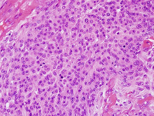
Washington University Experience | NEOPLASMS (MENINGIOMA) | Anaplastic | 13C3 Meningioma, anaplastic (Case 13) H&E 5.jpg
H&E stained sections of the resection material show a high grade meningioma with large, predominantly central, areas of geographic necrosis that comprise approximately 80% of the specimen. Mitotic figures are frequent, appearing at variable density that focally reaches 21/10HPF. Brain invasion is also identified focally. Sections of the dura show areas of tumor invasion.

